- Clinical Technology
- Adult Immunization
- Hepatology
- Pediatric Immunization
- Screening
- Psychiatry
- Allergy
- Women's Health
- Cardiology
- Pediatrics
- Dermatology
- Endocrinology
- Pain Management
- Gastroenterology
- Infectious Disease
- Obesity Medicine
- Rheumatology
- Nephrology
- Neurology
- Pulmonology
Can You Identify These Lesions?
Erythematous patches and papules on the knuckles; a slowly enlarging papule on a finger; a painful ulcer on the thumb; rough, thickened skin around the metacarpophalangeal joints; symmetric, soft, tan papules....

Figure 1

Figure 2
Case 1:
A 50-year-old man presents with erythematous patches and papules on the dorsa of his hands, mainly on the knuckles. Erythema and scale are noted on the proximal nail folds. Dermoscopic examination of the skin underneath the nail folds reveals telangiectasia.
The patient also has a persistent rash on the neck and anterior chest that worsens with sun exposure. For the past 3 months, he has had mild fatigue and vague weakness in the proximal extremities.
What is your take on these clinical findings?

Figure 1

Figure 2
Case 1:
The erythematous, slightly violaceous patches and papules on this patient's hands represent
Gottron sign and Gottron papules,
respectively. Both are commonly found on extensor surfaces. Gottron papules often develop on skin overlying dorsal phalangeal joints. The dermatologic findings, including the persistent neck rash, and the patient's history of fatigue and weakness suggest
dermatomyositis;
this autoimmune systemic disease can involve multiple organs.
The diagnosis can be confirmed by the results of laboratory studies, including measurement of antinuclear antibody, antihistidyl transfer RNA synthetase (anti-Jo-1), and creatine kinase, and by muscle biopsy. In this patient, an elevated creatine kinase level was confirmatory. Adults, especially those older than 50 years, with dermatomyositis need an age-appropriate workup for internal malignancy.
For this patient, treatment with oral prednisone resulted in moderate alleviation of his symptoms.

Case 2:
This skin-colored papule has been present on the index finger of a 35-year-old man for more than a year. The lesion is asymptomatic and has slowly enlarged.
What is this lesion? Is treatment necessary?

Case 2:
This is an
acquired digital fibrokeratoma,
or acral fibrokeratoma. The benign growth typically occurs as a solitary, soft or firm, skin-colored papule on a digit--usually a finger--in middle-aged adults. Although the pathogenesis is unclear, previous trauma may be a contributing factor. The differential diagnosis includes supernumerary digit, pyogenic granuloma, and wart.
Although the patient was reassured that the lesion was benign, he chose to have it removed by shave excision because of cosmetic concerns.

Figure 1

Figure 2
Case 3:
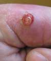
A 44-year-old man presents for evaluation of a painful, ulcerated nodule on the medial portion of the left thumb that has persisted for more than 6 months. The lesion bleeds frequently, especially following even mild trauma, and it has enlarged.
What is your clinical impression?

Figure 1

Figure 2
Case 3:
Based on the appearance of the nodule,
ulcerated pyogenic granuloma
was diagnosed. This benign lobular capillary hemangioma (or eruptive hemangioma) grows rapidly over a few weeks and presents as an erythematous papule or polyp. It occurs most commonly on the fingers, lips, and oral mucosa. Because of its friable and ulcerative nature, the lesion bleeds easily with mild trauma or rubbing. The cause is unknown.
In general, the diagnosis of pyogenic granuloma is based on the clinical history and examination findings. However, the lesion can mimic warts, hemangiomas, Kaposi sarcoma lesions, and even amelanotic melanomas. Biopsy is the only definitive method of diagnosis.
This patient's lesion was removed by shave excision, and the base of the tumor was cauterized. He applied bacitracin ointment to the area for 2 weeks until the wound healed. The lesion has not recurred.

Case 4:
A 39-year-old construction worker complains of rough, thickened skin on the dorsa of his hands and fingers, especially around the metacarpophalangeal joints.
What do you suspect?

Case 4:
These hardened plaques are
knuckle pads;
these benign, slow-growing tumors develop on the hands, especially on the metacarpal and proximal phalanges. Knuckle pads present as hyperpigmented or hypopigmented, fleshy, semifirm plaques. They are more likely to occur in middle-aged men, particularly those who perform hard labor.
This patient was reassured that the lesions were benign. For patients who are bothered by such lesions, postbathing application of urea (20% to 40%) or lactic acid (12.5%) creams or lotions may significantly reduce roughness and dryness.

Case 5:
During an evaluation for atopic dermatitis, soft, tan-colored papules are noted on the lateral aspect of the fifth finger on both hands of a 9-year-old boy. The papules are symmetric and asymptomatic. The patient's mother reports that the lesions have been present since birth.
What are these lesions?

Case 5:
These papules are
rudimentary supernumerary digits
(rudimentary polydactyly). These congenital accessory structures or appendages are typically located on the lateral surfaces of normal digits, most commonly on the lateral aspect of the fifth finger. They are symmetric and soft. In most patients, rudimentary supernumerary digits are asymptomatic; however, they can be painful, for instance, when there is trauma to the area.
In this patient, no treatment was necessary. His parents were informed that the lesions could be surgically excised in the future, if desired for cosmesis.
Atopic Dermatitis: The Pipeline and Clinical Approaches That Could Transform the Standard of Care
September 24th 2025Patient Care tapped the rich trove of research and expert perspectives from the Revolutionizing Atopic Dermatitis 2025 conference to create a snapshot of the AD care of the future.
