

- Clinical Technology
- Adult Immunization
- Hepatology
- Pediatric Immunization
- Screening
- Psychiatry
- Allergy
- Women's Health
- Cardiology
- Pediatrics
- Dermatology
- Endocrinology
- Pain Management
- Gastroenterology
- Infectious Disease
- Obesity Medicine
- Rheumatology
- Nephrology
- Neurology
- Pulmonology
Basal Cell Carcinoma on Arm of a 57-Year-Old Man
A small punch biopsy showed basal cell carcinoma with focal hypermelanosis. The underlying erythema and punctuate superficial dark black pigmentation strongly suggested a dysplastic nevus or even a melanoma.


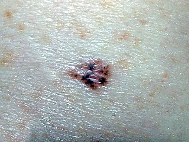
A 57-year-old man discovered a relatively small (1 cm), pigmented nodule of uncertain duration on his left arm during a cutaneous self-examination. The patient had a previous skin cancer.
Key point: The lesion displays an underlying erythema as well as punctuate superficial dark black pigmentation. While this strongly suggests a dysplastic nevus or even a melanoma, pigmented basal cell carcinoma can appear in this manner. A small punch biopsy disclosed basal cell carcinoma with focal hypermelanosis.
Treatment: The lesion was modestly sized and located on a low-risk skin region. Simple excision (with 5-mm borders) and primary closure was performed.
Note: The different diagnostic possibilities can be precise clinical mimics. Biopsy whenever doubt exists regarding the exact diagnosis.


