- Clinical Technology
- Adult Immunization
- Hepatology
- Pediatric Immunization
- Screening
- Psychiatry
- Allergy
- Women's Health
- Cardiology
- Pediatrics
- Dermatology
- Endocrinology
- Pain Management
- Gastroenterology
- Infectious Disease
- Obesity Medicine
- Rheumatology
- Nephrology
- Neurology
- Pulmonology
All Ears: Chondrodermatitis Nodularis Helicis and Verruca Vulgaris
Diagnostic challenge: Two case reports of easily treated and innocuous causes of lesions in the outer ear. Chondrodermatitis nodularis helicis is associated with long cellphone use. Verruca vulgaris is caused, like all other warts, by human papillomavirus.

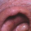
Chondrodermatitis nodularis helicis An 80-year-old man has had an asymptomatic, flesh-colored swelling on his right ear for 4 to 5 months. In the center is a 1-mm white scab pointing downward from the helix. At times, the patient shaves a white spicule that grows in this crusted area. He sleeps on his right side and does not use a cell phone.
The entire lesion was removed by elliptical excision under local anesthesia and sent for histopathologic examination. The diagnosis was chondrodermatitis nodularis helicis-a benign, usually solitary, chronically crusted nodule that develops most commonly on the superior helix of the right ear in men older than 50 years. In women, the lesion occurs less frequently and more commonly on the antihelix. The lesions can be painful.
The suspected cause of chondrodermatitis nodularis helicis is localized degeneration of dermal collagen with its subsequent partial extrusion through a central ulceration. Long-term cell phone use is associated with the development of this lesion at the point of pressure on the ear.1 Other possible causative factors include minor trauma, pressure during sleep,2 poor vascularity, and solar damage.3 Systemic sclerosis and dermatomyositis are rare associations.4,5 Perforation of the infundibular portion of a hair follicle with extrusion of its contents into the dermis is another suggested cause.6,7
Treatment options include excision, cryotherapy or curettage, and cautery. A recurrence rate of up to 20% has been reported.3 This patient's ear healed without complication. The lesion has not recurred.

Verruca Vulgaris
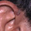
This tan, 7x3x5-mm, papillary lesion with a broad, slightly elevated, eccentric base has been present for several months on the right anterior helix of a 24-year-old man. It is asymptomatic.
The lesion was removed by shave excision in the office under local anesthesia, and its base was desiccated and curetted. Histopathologic examination confirmed the clinical diagnosis of verruca vulgaris.
Common warts may occur on any skin surface- although they most frequently occur on the hand and fingers--and are usually few in number. These benign lesions are caused by human papillomaviruses (HPVs). More than 100 types of HPVs exist, with new types discovered each year. The clinical manifestations (sites) of warts vary depending on the HPV type. The common wart is associated with HPV types 2, 4, and 29.1
Treatment of warts depends on their size and location. Options include cryotherapy, electrodesiccation and curettage, topical keratolytic agents, immunomodulator drugs, vitamin A, laser, chemotherapy, formalin, intralesional bleomycin, and duct tape occlusion. Suggestive therapy may work for some patients.
After this patient's lesion was removed, antibiotic ointment was applied. The ear healed with excellent cosmetic results.
See also:
All Ears: Auricular Seroma and Pyogenic Granuloma
All Ears: Seborrheic Keratosis and Actinic Keratosis
References:
REFERENCES:
1.
Elgart ML. Cell phone chondrodermatitis.
Arch Dermatol.
2000;136:1568.
2.
Dean E, Bernhard JD. Bilateral chondrodermatitis nodularis antihelicis. An unusual complication of cardiac pacemaker insertion.
Int J Dermatol.
1988; 27:122.
3.
Bard JW. Chondrodermatitis nodularis chronica helicis.
Dermatologica.
1981;163:376-384.
4.
Bottomley WW, Goodfield MDJ. Chondrodermatitis nodularis helicis occurring with systemic scleroissi--an under-reported association.
Clin Exp Dermatol.
1994;19:219-220.
5.
Sasaki T, Nichizawa H, Sugita Y. Chondrodermatitis nodularis helicis in childhood dermatomyositis.
Br J Dermatol.
1999;141:363-365.
6.
Hurwitz RM. Painful papule of the ear: a follicular disorder.
J Dermatol Surg Oncol.
1987;13:270-274.
7.
Hurwitz RM. Pseudocarcinomatous or infundibular hyperplasia.
Am J Dermatopathol.
1989; 11:189-191.
