- Clinical Technology
- Adult Immunization
- Hepatology
- Pediatric Immunization
- Screening
- Psychiatry
- Allergy
- Women's Health
- Cardiology
- Pediatrics
- Dermatology
- Endocrinology
- Pain Management
- Gastroenterology
- Infectious Disease
- Obesity Medicine
- Rheumatology
- Nephrology
- Neurology
- Pulmonology
All Ears: Auricular Seroma and Pyogenic Granuloma
These innocuous lesions of the outer ear may arise spontaneously or after trauma or surgery. Both auricular seroma and pyogenic granuloma usually resolve satisfactorily after minor surgery, though they may recur.

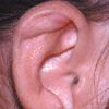
Auricular SeromaA 35-year-old woman is concerned about a 1-cm swelling that suddenly appeared in the superior crus of the right antihelix. The lesion is fluctuant and asymptomatic; no tenderness or warmth is felt on palpation. She denies trauma to the area.
Aspiration of the lesion with a large bore needle yielded 1.5 mL of clear serous fluid. It was felt the lesion represented an auricular seroma, which probably resulted from the shearing force of her head against her pillow during sleep.
Generally, seromas tend to develop in incisions after surgery. Auricular seromas are rare, but they should be included in the differential diagnosis of an asymptomatic, noninflammatory swelling of the ear. The lesions may drain spontaneously. In this patient, simple aspiration was curative.

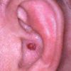
Pyogenic granulomaThis asymptomatic, 0.5-cm, red berry-like lesion in the concha of a 39-year-old man's left ear has been gradually enlarging for 3 months.
A benign hemangioma of mucous membranes and skin, pyogenic granuloma occurs most commonly on the gingiva, lips, fingers, and face and less commonly on the trunk, arms, legs, and conjunctiva. Most are polypoid or pedunculated; they are less commonly sessile. The base is often surrounded by a ring of fine scale.1
Pyogenic granulomas evolve rapidly over weeks, frequently become ulcerated, and bleed easily. The lesions can develop at any age, in both sexes, and in a nevus flammeus or spider angioma. Occasionally, more than 1 lesion is present. Most pyogenic granulomas occur spontaneously; however, they may follow trauma, retinoid therapy, injury (such as an insect bite, burn, or scald), or cryotherapy.
Spontaneous resolution of a pyogenic granuloma is uncommon, except in the case of epulis gravidarum (pyogenic granuloma of the gingiva that develops during pregnancy). Treatment includes excision or desiccation and thorough curettage. If any tissue remains, recurrence is likely.
In this patient, the lesion was removed via shave excision, treated with light curettage, and cauterized with ferric subsulfate solution. Topical mupirocin ointment was applied until the ear healed.
See also:
All Ears: Chondrodermatitis Nodularis Helicisand Verruca Vulgaris
All Ears: Seborrheic Keratosis and Actinic Keratosis
References:
REFERENCE:
1.
Elgart ML. Cell phone chondrodermatitis.
Arch Dermatol.
2000;136:1568.
