- Clinical Technology
- Adult Immunization
- Hepatology
- Pediatric Immunization
- Screening
- Psychiatry
- Allergy
- Women's Health
- Cardiology
- Pediatrics
- Dermatology
- Endocrinology
- Pain Management
- Gastroenterology
- Infectious Disease
- Obesity Medicine
- Rheumatology
- Nephrology
- Neurology
- Pulmonology
Addison's Disease
Primary adrenal insufficiency or Addison’s disease, is the result of destruction of the adrenal cortex.
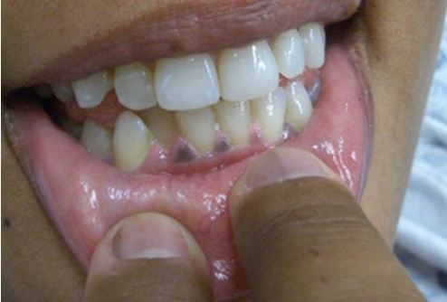
Fig 2. Gum hyperpigmentation in Addison's disease.

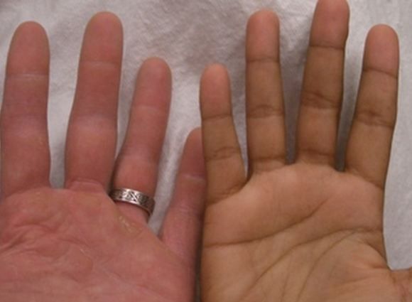
Fig 3. Palmar crease hyperpigmentation in Addison's disease.

A 31-year-old Caucasian woman presents to her doctor complaining of intermittent episodes of weakness, upper abdominal discomfort, and nausea that last several days at a time and have been occurring for the past 6 months. She also states that her skin is getting a lot darker, although she has not been in the sun more than usual. She denies any fever, diarrhea, vomiting, polyuria, bleeding, or irregular menstrual periods. She is on thyroid replacement therapy for hypothyroidism but has no other medical conditions and does not take other medications.
On examination she is alert, tan, and in no acute distress. Vital signs are normal except for a BP of 84/56 mm Hg but she denies feeling light headed. She appears well hydrated. Her abdomen is not tender. The exam is otherwise unremarkable except for her notably dark complexion. In response to a request for an old photograph, she provides her driver’s license; compared to that picture, her skin appears much darker. The image of her fingers (Figure 1, below) was taken on the day of the visit.
[[{"type":"media","view_mode":"media_crop","fid":"42023","attributes":{"alt":"Figure 1. ","class":"media-image","id":"media_crop_9286049616314","media_crop_h":"0","media_crop_image_style":"-1","media_crop_instance":"4510","media_crop_rotate":"0","media_crop_scale_h":"0","media_crop_scale_w":"0","media_crop_w":"0","media_crop_x":"0","media_crop_y":"0","style":"height: 317px; width: 435px;","title":" ","typeof":"foaf:Image"}}]]
Based on the history, what is the most likely diagnosis?
What is the appropriate treatment?
Answer: Addison’s disease (aka, primary adrenal insufficiency)
Although there are many conditions that can cause nausea and abdominal pain, there are few conditions that cause global hyperpigmentation. The combination is most consistent with primary adrenal insufficiency (Addison’s disease). At right, Figure 2 shows gum hyperpigmentation and Figure 3 highlighs palmar crease hyperpigmentation (author’s hand shown on the left for comparison; click on images to enlarge).
Discussion
Adrenal insufficiency is most commonly secondary to pituitary disease (secondary adrenal insufficiency) wherein the pituitary gland secretes less adrenocorticotropic hormone (ACTH) which, in turn, affects glucocorticoid production, but not mineralocorticoid production. Symptoms tend to be mostly gastrointestinal with nausea, abdominal pain, and sometimes vomiting but rarely diarrhea. Examination may be normal except in severe cases.
Basic testing may reveal low glucose but if the diagnosis is suspected a cosyntropin stimulation test should be done: 250 µg of ACTH are administered via IV and cortisol levels are measured at 30 and 60 minutes. Initial treatment involves IV saline with dextrose and corticosteroid replacement with Solu-Cortef or Decadron. Long term treatment involves chronic steroid replacement.
Primary adrenal insufficiency or Addison’s disease, is the result of destruction of the adrenal cortex. It is less common than secondary adrenal insufficiency but more severe and pronounced because mineralocorticoids are also affected, causing dehydration and electrolyte abnormalities. In addition, feedback mechanisms result in increased pituitary production of ACTH in Addison's disease. ACTH also stimulates melanocytes, leading to the signature hyperpigmentation that affects not only sun-exposed skin but also the palms and even the gums (Figures 2 and 3). Symptoms are otherwise similar to secondary adrenal insufficiency but are often more pronounced with more hypotension. Initial treatment is similar, including IV fluids, supportive care and steroid replacement. Long term both glucocorticoids and mineralocorticoids will require replacement.
{C}
Secondary Adrenal Insufficiency (more common than Addison’s Disease):
↓glucose. K & Na both normal, calcium and eosinophil count should be normal too
Cosyntropin stimulation test: Cortisol level <20 at 30 & 60 minutes after ACTH 250µg
D5NS, Solu-Cortef 100mg IV or Decadron 6mg (won’t ruin stimulation test) (vasopressors)
Click Image for Details
