- Clinical Technology
- Adult Immunization
- Hepatology
- Pediatric Immunization
- Screening
- Psychiatry
- Allergy
- Women's Health
- Cardiology
- Pediatrics
- Dermatology
- Endocrinology
- Pain Management
- Gastroenterology
- Infectious Disease
- Obesity Medicine
- Rheumatology
- Nephrology
- Neurology
- Pulmonology
Acute Ankle Pain in a Runner
A healthy 23-year-old woman, who is a longtime runner, calls your office the day after shetripped on a tree root while running in the woods and twisted her right ankle. She noted immediatepain on the lateral side of the ankle but did not hear or feel a pop. She was able tobear weight and walked out of the woods. However, as she walked, the ankle became morepainful and began to swell. When she reachedhome, she applied a heating pad and restedthe ankle. The next morning she noted increasedswelling and moderate discomfortwhile walking.
PATIENT PROFILE:
A healthy 23-year-old woman, who is a longtime runner, calls your office the day after she tripped on a tree root while running in the woods and twisted her right ankle. She noted immediate pain on the lateral side of the ankle but did not hear or feel a pop. She was able to bear weight and walked out of the woods. However, as she walked, the ankle became more painful and began to swell. When she reached home, she applied a heating pad and rested the ankle. The next morning she noted increased swelling and moderate discomfort while walking.
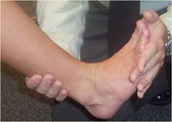
She now is concerned that she will be unable to run. She has no history of past ankle injuries, and she has no known medical problems or allergies.
WHICH TWO OF THE FOLLOWING WOULD YOU DO NOW?
A. Advise the patient to continue to use the heating pad.
B. Tell her to stop using heat and switch to ice.
C. Order a radiograph of the ankle.
D. Have the patient elevate the right leg and compress the ankle with an Ace bandage.
(Answer on next page.)THE CONSULTANT’S CHOICE
The history suggests that the patient has probably stretched or torn one of the lateral ankle ligaments. The anterior talar fibular ligament and the calcaneofibular ligament are the most commonly injured lateral ankle ligaments.
The immediate swelling indicates bleeding, which was likely increased by the patient’s use of a heating pad. Heat (choice A)-because it causes vasodilatation- increases bleeding, swelling, and inflammation and thus is not appropriate at this point. However, heat is helpful in the later rehabilitation stages.
It is vital to make every effort to reduce the swelling, because it decreases range of motion. Rehabilitation time is increased with decreased range of motion.
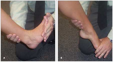
Figure 1 –These photographs show how to test for dorsiflexion (A) and plantar flexion (B) in a patient’s ankle.
Ice (choice B) reduces pain and swelling. It also decreases metabolism and limits hypoxic injury.1 A sprained ankle should be cooled for 20 minutes every 3 to 4 hours during the first 48 hours. The ice can be placed in a plastic bag and applied to the ankle over a thin cloth. Compression and elevation (choice D) also help decrease swelling. Rest, ice, compression, and elevation together form the commonly used acronym “RICE.” 1
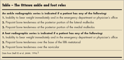
A radiograph (choice C) is not indicated at this point. The Ottawa ankle rules2 are a simple and reliable tool for deciding when to obtain a radiograph in an ankle injury (Table). A physical examination is necessary in order to apply all aspects of the rules. However, one of the key points in the Ottawa ankle rules is that the ability to bear weight usually means a radiograph is not indicated. This patient is able to walk and bear weight on her ankle.
The patient follows your recommendations, then comes to your office the next day for evaluation and further treatment. Examination of the ankle reveals swelling and diffuse tenderness over the right lateral malleolus. Dorsiflexion (Figure 1) is limited to 10 degrees in the right foot compared to 20 degrees in the left (normal dorsiflexion is 15 to 25 degrees). Plantar flexion (see Figure 1) is equal bilaterally at 45 degrees (normal plantar flexion ranges between 40 and 55 degrees).
WHAT WOULD YOU DO NOW?A. Perform a squeeze test.
B. Perform a talar tilt test.
C. Perform an anterior drawer test.
D. Palpate over the base of the fifth metatarsal.
E. Palpate for tenderness over the posterior portions of the lateral and medial malleolus.
F. All of the above.
(Answer on next page.)THE CONSULTANT’S CHOICE
Quality care for an ankle sprain includes the performance in the physical examination of all of the above tests (choice F). These tests help identify which ligaments- if any-are damaged and help assess the extent of the damage. A thorough examination also assists with the selection of therapy and determination of the patient’s prognosis.
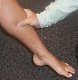
Figure 2 –To perform the squeeze test, compress the midleg from posterior lateral to anterior medial. The squeeze test is used to identify disruption of the tibiofibular syndesmosis (the interosseus membrane between the tibia and fibula).
The squeeze test is used to identify disruption of the tibiofibular syndesmosis (the interosseus membrane between the tibia and fibula). To perform the test, compress the midleg from posterior lateral to anterior medial (Figure 2). This test separates the fibula and tibia and will produce pain in the lower ankle in the tibiofibular syndesmosis if that membrane has been disrupted.
The talar tilt test is performed with the patient seated. Secure the lower leg with one hand and grasp the forefoot with the opposite hand. Apply an inversion force to produce a talar tilt. Perform the test with the ankle in both the neutral position and in plantar flexion (Figure 3). It is important to perform the talar tilt test in both ankles and compare the results. The calcaneofibular ligament (CFL) stabilizes the ankle in the neutral position; thus, stress in this position tests the integrity of the CFL. The anterior talofibular ligament (ATFL) stabilizes the ankle in plantar flexion; thus, stressing a plantar-flexed ankle tests the integrity of the ATFL. Bear in mind that, in general, the ankle is less stable in plantar flexion because of the configuration of the talus: the anterior margin is wider than the posterior margin by an average of 2.4 mm. This configuration imparts stability to the ankle joint during dorsiflexion and relative instability during plantar flexion.
The anterior drawer test also assesses the integrity of the ATFL. While the patient is seated, grasp the lower leg with one hand and the forefoot with the other. Then, apply an anterior force to produce forward translation ( Figure 4). Perform the test with the ankle in both the neutral position and in plantar flexion. Compare results in the 2 ankles. A few millimeters of translation is normal. The test is more reliable in chronic instability than in acute instability because the inhibition caused by the pain of an acute sprain can lead to a falsenegative result. Therefore, when the result is negative in an acute ankle sprain, repeat the test after the pain subsides.
Palpation over the base of the fifth metatarsal tests for a fracture that has been caused by an avulsion of the peroneus brevis muscle. Palpation for tenderness over the posterior portions of the lateral and medial malleolus tests for fractures in these bones. Palpation for pain in these areas is one of the prerequisites for application of the Ottawa ankle rules (see Table).
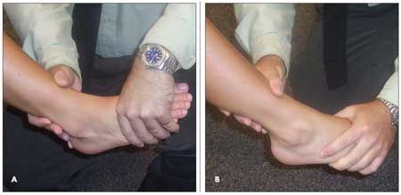
Figure 3 –Perform the talar tilt test with the ankle in both the neutral position (A) and in plantar flexion (B). With the patient seated, secure the lower leg with one hand and grasp the forefoot with the opposite hand. Apply an inversion force to produce a talar tilt.
Examination of this patient’s ankle reveals swelling and some ecchymosis over the lateral malleolus. The anterior and medial portions of the lateral malleolus are tender, but the posterior portion of the lateral malleolus is not. The medial malleolus is not tender at all. Results of the squeeze and anterior drawer tests are negative. The talar tilt test produces pain with the ankle in plantar flexion but no instability. Palpation of the base of the fifth metatarsal reveals no tenderness.
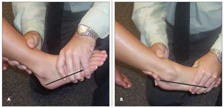
Figure 4 –The anterior drawer test shown here assesses the integrity of the anterior talofibular ligament. With the patient seated, grasp the lower leg with one hand and the forefoot with the other. Then, apply an anterior force to produce forward translation (arrows). Perform the test with the ankle in both the neutral position (A) and in plantar flexion (B).
WHAT WOULD YOU NOT DO NOW?
A.
Obtain a radiograph of the ankle.
B.
Suggest that the patient wear an ankle brace for 3 to 4 weeks.
C.
Recommend that she start a rehabilitation program.
D.
Prescribe an NSAID.
(Answer on next page.)
THE CONSULTANT’S CHOICE
A radiograph of the ankle (choice
A
) is not indicated. Application of the Ottawa ankle rules provides the rationale for this determination (see
Table
). There is no tenderness in the specified areas, and the patient is able to bear weight.
Stretching or tearing of the lateral ankle ligaments leads to inversion instability. An ankle brace (choice B) improves stability and enables a patient to return to work or play sooner. However, a brace is only a temporary measure and should not substitute for a rehabilitation program. The same caution applies to the use of NSAIDs (choice D). These agents can be started immediately after an injury and continued for up to 10 days. They are perfectly acceptable as an adjunct to a rehabilitation program. Remind patients to take NSAIDs with meals, and do not use these agents in patients with significant hypertension, peptic ulcer/gastroesophageal reflux disease, or renal disease.
A rehabilitation program (choice C) can be started within hours of an injury unless the ligament is completely ruptured. Prolonged immobilization is not recommended; it leads to decreased range of motion and lengthy recovery.
Many acute ankle injuries become chronic injuries because of a lack of proper rehabilitation. The 4 components of rehabilitation are:
•Range of motion exercises.
•Progressive muscle strengthening.
•Proprioceptive training.
•Activity-specific training.
This patient has already lost 10 degrees of dorsiflexion; thus, she needs to start dorsiflexion exercises now. In general, movements that encourage dorsiflexion and plantar flexion, such as writing the alphabet with the foot, can be started early on. After 48 hours, have patients begin stretching the Achilles tendon with a towel to increase dorsiflexion. Direct them, while seated, to wrap a towel around their toes and use this to pull the toes toward their head.
Have patients continue the RICE routine as long as pain and swelling persist. Once the pain has diminished, a more aggressive program is indicated. Strengthening of weakened muscles through the use of resistance exercises and toe raises is essential to rapid recovery.
When the patient achieves full weight bearing without pain, begin proprioceptive training to aid in the recovery of balance and postural control. Patients can be referred to a trainer or physical therapist for this training.
Activity-specific training is most important for patients involved in athletic activities, such as basketball and football, that require quick changes of direction. Exercises that develop agility with quick directional changes can be taught by the physician or by a physical therapist or trainer.
This patient received a prescription for an NSAID, and she was taught some initial exercises to stretch and strengthen her ankle ligaments. After 2 weeks, she returned to running but was unable to complete her runs without lateral ankle pain. She then was referred to a physical therapist; after 2 months she returned to her former running routine.
References:
REFERENCES:
1.
Knight KL. Initial care of acute injuries: the RICE technique. In:
Cryotherapyin Sport Injury Management.
Champaign, Ill: Human Kinetics; 1995:209-215.
2.
Stiell IG, McKnight RD, Greenberg GH, et al. Implementation of the Ottawaankle rules.
JAMA.
1994;271:827-832.
AI-Powered Diabetes Prevention Program Intervention Matches Human Coaching in Landmark Trial
October 27th 2025After 1 year of coaching by the AI app or a DPP-associated coach, reductions in weight and HbA1c and increases in physical activity were equal, an outcome with great promise for scalability.
