- Clinical Technology
- Adult Immunization
- Hepatology
- Pediatric Immunization
- Screening
- Psychiatry
- Allergy
- Women's Health
- Cardiology
- Pediatrics
- Dermatology
- Endocrinology
- Pain Management
- Gastroenterology
- Infectious Disease
- Obesity Medicine
- Rheumatology
- Nephrology
- Neurology
- Pulmonology
16-Year-Old Camper With Tibial Pain
A 16-year-old boy complains of right lower leg pain that began after his first week at a summer basketball conditioning camp. Before he left for the camp, he was jogging off and on, averaging a few miles a week.
A 16-year-old boy complains of right lower leg pain that began 2 weeks earlier, after his first week at a summer basketball conditioning camp. Before he left for the camp, he was jogging off and on, averaging a few miles a week. At camp he began running 7 miles a day and doing sprints 3 times a week.

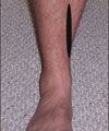
The leg pain initially began at the end of a run and quickly disappeared. It now is present as soon as he starts his run, and after 15 to 20 minutes it is so intense that he cannot continue. The pain persists for 1 to 2 hours after a run. It is located on the posteromedial aspect of the right tibia, covering an area that starts about 1 inch above the medial malleolus and extends upward about 4 inches (Figure 1). He does not note any numbness or dragging of his right foot. He is in good health and has never had a similar problem.
Physical examination reveals no gross difference in appearance between the 2 lower extremities. The patient pronates when he walks. Both plantar flexion against resistance and standing on tiptoes reproduce the pain in his right leg. The result of a tuning fork test is negative, and he exhibits no pinpoint tenderness. He has no neurologic or vascular deficit. His shoes are 2 years old and were used by his brother for a full season. They provide minimal support medially, and the soles are significantly worn.
Which diagnosis is most likely?
A. Tibial stress fracture.
B. Compartment syndrome.
C. Posterior tibial tendonitis.
D. Medial tibial stress syndrome (MTSS).
THE CONSULTANT'S CHOICE
Given the patient's age, the lack of acute trauma, the gradual onset of symptoms, and the recent initiation of intense physical activity, this is most likely an overuse injury. All 4 choices represent overuse injuries that can occur in runners.


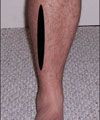
The location of the pain and the lack of symptoms of neurologic or vascular compromise (such as numbness or footdrop) make compartment syndrome (B) less likely. Compartment syndrome associated with running is usually located in the anterior or lateral portion of the leg (Figure 2).
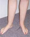
Posterior tibial tendinitis or tendinopathy (C) follows the path of the posterior tibial tendon, posterior to the medial malleolus. The pain associated with this injury is usually located behind the medial malleolus (Figure 3) and is typically accompanied by swelling. The lack of swelling and of pain in the location of the medial malleolus makes this diagnosis unlikely.
The location of the pain over the area of the posterior medial tibia is characteristic of both stress fractures (A) and MTSS (D). Although stress fractures are usually located in the medial posterior region of the tibia, the area of pain is typically much smaller than that seen in this patient.
The tuning fork test can help rule out a stress fracture. With the fork vibrating, place the blunt end superior to the area of pain. Feeling increased pain in the area previously described as painful is considered a positive result. However, this test is of more diagnostic value when the result is negative (ie, when there is no increase in perceived pain). The negative result on a tuning fork test and the lack of pinpoint tenderness both make a stress fracture unlikely here.
Pronation-which this patient exhibits-predisposes the posterior musculature of the lower leg to pull excessively on the tibia. This pull or stretch causes a periosteal reaction on the tibia at its posterior medial border. Because this boy's shoes provide minimal medial support, they aggravate the medial tibial stress. These clues, in conjunction with the location of the pain, point to MTSS (also called "shin splint syndrome").
Which study would you order now?
A. Plain radiograph.
B. MRI scan.
C. Measurement of compartment pressures.
D. No further testing.
THE CONSULTANT'S CHOICE
Compartment pressures (C) are not indicated because compartment syndrome seems unlikely. An MRI scan (B) is overkill at this point: it is not indicated and thus would add considerable unnecessary expense. Many clinicians would order a plain radiograph as a precaution, to check for stress fracture; however, this, too, is an additional expense. If the patient does not respond to treatment, a radiograph would then be indicated. At this time, however, no further testing is recommended.

Pathophysiology of MTSS. This overuse syndrome is caused by a mismatch between overload and recovery. Repetitive overload on tissues that are not able to adapt to new or increased demands leads to tissue breakdown. Specifically, MTSS results from traction of the posterior tibial, flexor digitorum longus, and soleus muscles on the periosteum of the tibia (Figure 4). The traction produces an overload stress, which leads to an inflammatory response in the tibial periosteum (periostitis).
Clinical findings. The pain of MTSS is located over the posteromedial tibia beginning just above the medial malleolus and extending superiorly for 3 to 5 inches (see Figure 1). It is activity-related, and chronic neurologic symptoms, such as numbness and footdrop, are usually absent. Diffuse pain and tenderness along the posteromedial border of the tibia that are aggravated by plantar flexion are characteristic findings. Pronation of one or both feet may be present.
Contributing factors. Multiple factors can contribute to or increase the risk of overload. A prior injury or lack of conditioning may lead to muscle imbalance, inflexibility, weakness, and instability. Other factors that can contribute to the development of MTSS include poor exercise technique, improper equipment, and changes in the duration or frequency of activity (training errors). Some persons seem to be more vulnerable to MTSS than others. This boy was not well conditioned when he began camp, his equipment (footwear) was faulty, and he pronated. His anatomy predisposed him to overload his posterior leg musculature, and his lack of conditioning contributed muscle weakness and lack of flexibility.
Which treatment would you recommend?
A. A course of NSAIDs.
B. An orthosis to correct the patient's pronation.
C. Calf strengthening exercises.
D. Cross training on a stationary bicycle.
E. All of the above.
THE CONSULTANT'S CHOICE
Treatment of MTSS is conservative. Running needs to be stopped for a short period, but alternative activities, such as swimming or use of a stationary bicycle, can be substituted. Additional treatment options include NSAIDs to reduce inflammation, orthoses to correct pronation, and exercises to strengthen the calf muscles.
Outcome of this case. This patient's treament included all of these options. He began a 10-day course of ibuprofen, 600 mg tid. An orthosis was prescribed to correct his pronation. (He also obtained new shoes that had strong medial support.) He refrained from competitive basketball for 2 weeks and from weight-bearing exercise for 1 week. He used a stationary bicycle for 1 week; then he began to alternate walking/jogging with riding the stationary bike. The camp's trainer oversaw a program of gradually increasing activity. He did very well, and within 4 weeks had returned to full play.
