- Clinical Technology
- Adult Immunization
- Hepatology
- Pediatric Immunization
- Screening
- Psychiatry
- Allergy
- Women's Health
- Cardiology
- Pediatrics
- Dermatology
- Endocrinology
- Pain Management
- Gastroenterology
- Infectious Disease
- Obesity Medicine
- Rheumatology
- Nephrology
- Neurology
- Pulmonology
ECG Challenge: Neck Pain and a Mystery Fall
Severe pain from a pinched nerve in his neck could have caused this 64-year-old man to fall to the floor from his bed - but he's not sure. Does the ECG offer a clue?
Figure 1. (Please click to enlarge)

Figure 2. (Please click to enlarge)

Figure 3. (Please click to enlarge)

History: A 64-year-old man with a history of hypertension and chronic neck pain is brought to the ED by ambulance for neck pain, possible syncope, and a forehead laceration. Medics state there was minimal blood loss and initial vital signs, glucose, and field ECG all were normal. The patient states that for the past few days he has not been sleeping well due to a pinched nerve in his neck with pain radiating into his left shoulder. He states he was sitting on the edge of his bed in pain and believes he fell asleep and toppled to the ground. He woke up on the floor with a forehead laceration.
He does not think he blacked out but is not sure. He denies any headache, vomiting, chest pain, palpitations, or other complaints. He states his shoulder and neck are both mysteriously less painful after the fall than before.
Examination: Vital signs were normal. Physical examination was normal except for a 5-cm forehead laceration.
Initial Concerns: Syncope, cervical radiculopathy, ACS mimicking pre-existing musculoskeletal condition.
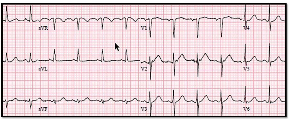
Testing: See ECG tracing in Figure 1, above (please click on image to enlarge).
Questions1. What does the case image show?
2. What should you do next?
Please click here for answers and next question.
Answers1. What does the case ECG image show? Not much. There are flat T waves in aVL and the upslope of the T-waves in V2 and V3 are a bit straighter than normal. These are nonspecific findings
2. What should you do next? Repeat the EKG.
The ECG was unchanged. On the way to nuclear medicine for CT scan of his head, the nurse calls to let you know that the patient's left shoulder pain suddenly got a lot worse and that he looks uncomfortable. She wants to know if they should finish the scan or bring him back for pain medication.
3. What should you tell the nurse?
Please click here for answer and discussion.
Answer
3. What should you tell the nurse? Tell the nurse to bring the patient back immediately so you can repeat the ECG.
Discussion
It is important to be vigilant for atypical presentations of acute coronary syndrome (ACS) and to have a low threshold for ordering serial ECGs, especially when pain has recurred or changed or when pain is ongoing and of recent onset. ACS can mimic a pre-existing musculoskeletal condition and may even have a tendency to do so. Be particularly suspicious for ACS in patients who tell you the pain feels like their usual pinched nerve or rotator cuff tendonitis, especially when the pain is in any way exertional.
Also be vigilant for ongoing or changing symptoms in a patient with an initially normal ECG, especially if that ECG was performed when the patient was asymptomatic or within the first 2 or so hours of the pain. Just as an initial troponin level may be falsely normal in ACS, especially early on, so, too, may the ECG. This occurs in about 15% of cases of ST-elevation MIs. The first question I usually ask a patient with possible ACS is “Are you still having discomfort right now?” and if the answer is yes, “When did the current episode start?”
There are guidelines that recommend serial ECGs every 15 to 30 minutes for the first hour of pain if that pain is ongoing. To be safe, another ECG should be performed about 2 hours after pain onset to avoid missing ST elevation that is delayed in onset.
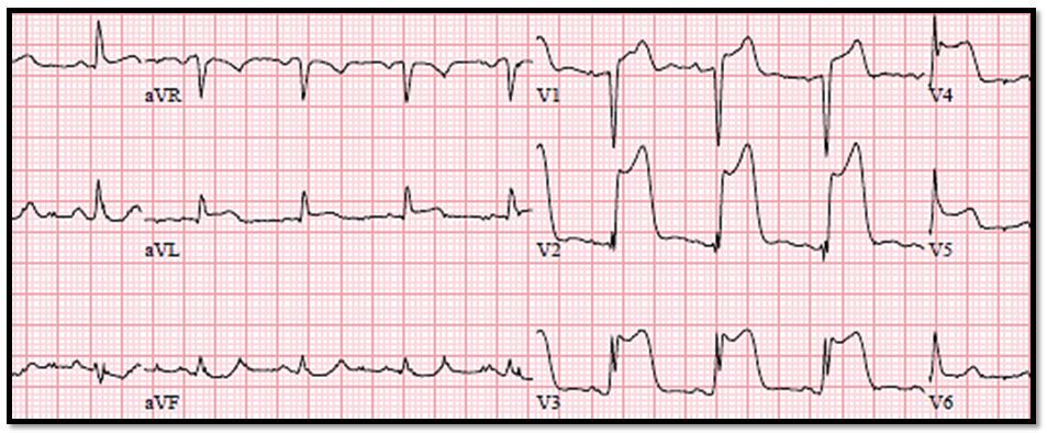
Case conclusion: Tracing from the patient's repeat ECG is shown in Figure 2, above. The patient was taken to the cath lab, where he had angioplasty and stenting of a completely occluded LAD.
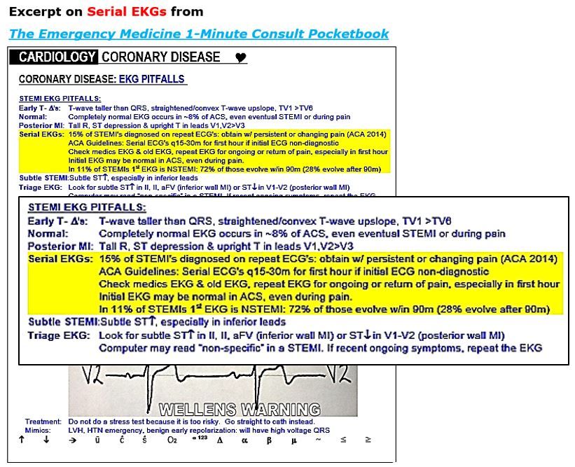
For more details on ECG pitfalls, click on page shot in Figure 3 above and view highlighted area.
