- Clinical Technology
- Adult Immunization
- Hepatology
- Pediatric Immunization
- Screening
- Psychiatry
- Allergy
- Women's Health
- Cardiology
- Pediatrics
- Dermatology
- Endocrinology
- Pain Management
- Gastroenterology
- Infectious Disease
- Obesity Medicine
- Rheumatology
- Nephrology
- Neurology
- Pulmonology
Acute Tenesmus, Night Sweats, and Generalized Weakness
A 62-year-old man is transported to the ED after becoming incontinent in the middle of the night. He has a history of HCV. What do test results suggest?
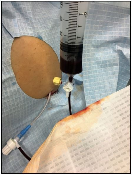
Image. Diagnostic paracentesis (please click to enlarge)

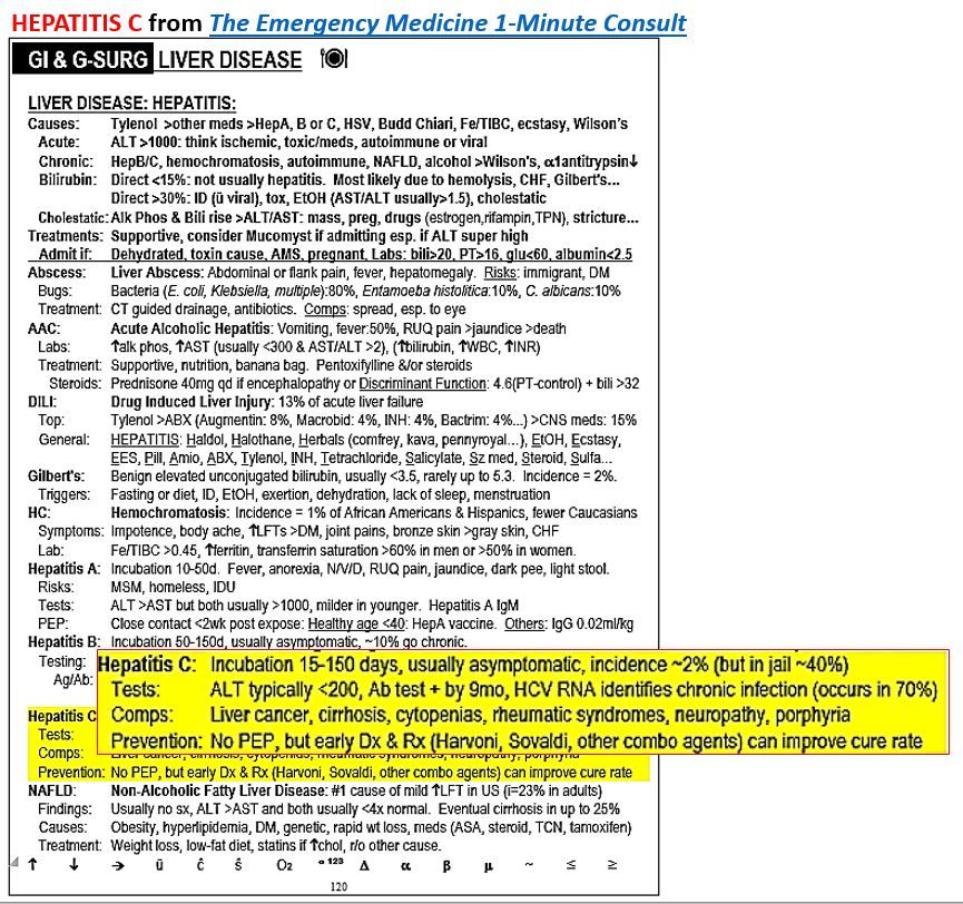
Figure. (Please click to enlarge)

Patient history
A 62-year-old man with a history of hypertension and hepatitis C cirrhosis is brought to the emergency department by paramedics for near syncope followed by abdominal pain. He states he was fine when he went to sleep, but then woke at 1 AM with tenesmus, night sweats, chills, and severe generalized weakness.
He was too weak to even get out of bed to go to the toilet and ended up incontinent of stool in his bed after which he called 911. En route to the ED he developed generalized abdominal pain. He denies any chest pain, vomiting, melena, or other complaints.
Examination
Vital signs were notable for blood pressure in the 70s and a temperature of 93.2°F. Pulse rate and respiratory rate were both normal.
Physical examination was notable for rigors, cool clammy skin, epigastric tenderness, positive fluid wave test, and guaiac-positive tan-colored stool that was all over his legs and back.
Initial concerns
- Sepsis
- GI bleed
- Spontaneous bacterial peritonitis (SBP)
Laboratory resultsChemistry:Lactate 9.2 mmol/L, INR 2.5
CBC: WBC normal, hemoglobin 6.5 g/dL, platelets 49
Paracentesis:a small volume diagnostic paracentesis was done to check for SBP. The safety-tip trocar was introduced into a large pocket of free fluid with ultrasound guidance. The first and only syringe of fluid aspirated is shown in image above (please click on image to enlarge).
Questions 1. What does the case image show? 2. What should you do next?
Please click here for answers, diagnosis, and discussion.
Answers
1. What does the case image show? Frank blood
2. What should you do next? Leave the catheter in place. Call interventional radiology (IR) for angioembolization. The bleeding is most likely due to rupture of a hepatocellular carcinoma secondary to hepatitis C.
For details: See yellow highlighted area of sample page in the Figure at right (please click on image to enlarge).
Discussion
Hepatitis C is currently considered an epidemic in the US, but with a very delayed clinical presentation. The incidence in the US is estimated to be ~2%, although it is about 20 times higher in those with certain risk factors, most notably a history of injection drug use or incarceration. Hepatitis C can be spread sexually, but this is rare and far less common than with HIV or hepatitis B.
Hepatitis C is usually asymptomatic for years to decades and most patients do not discover they have it until they develop complications from chronic infection or liver functions tests are found to be elevated during routine laboratory testing. The ALT will typically be the highest value, but usually remains below 200 IU/L. For more specific information on incubation period and timing of other test result abnormalities click on the sample page in the Figure, then focus on the highlighted area.
Complications of hepatitis C are protean and include hepatocellular carcinoma, cirrhosis, neuropathy, and porphyria to name a few. Carcinomas in particular have a very poor prognosis and occasionally rupture causing acute life-threatening internal bleeding, as in this case. Treatment of such a rupture is usually with angioembolization, but surgery may also be required.
Remember this: stool incontinence, unless specifically caused by profuse diarrhea, should always be considered a very concerning finding in a previously independent adult
Conclusion
Paracentesis was stopped and the pigtail catheter was left in place. Surgery and IR were consulted. A CT of the abdomen was performed and showed large volume ascites with hyperdense blood layering dependently within the pelvis as well as a liver mass suspicious for hepatocellular carcinoma. The patient was taken to interventional radiology for embolization.
Additional information is availabe at: The Emergency Medicine 1-Minute Consult pocket book
Clinical Tips for Using Antibiotics and Corticosteroids in IBD
January 5th 2013The goals of therapy for patients with inflammatory bowel disorder include inducing and maintaining a steroid-free remission, preventing and treating the complications of the disease, minimizing treatment toxicity, achieving mucosal healing, and enhancing quality of life.
