- Clinical Technology
- Adult Immunization
- Hepatology
- Pediatric Immunization
- Screening
- Psychiatry
- Allergy
- Women's Health
- Cardiology
- Pediatrics
- Dermatology
- Endocrinology
- Pain Management
- Gastroenterology
- Infectious Disease
- Obesity Medicine
- Rheumatology
- Nephrology
- Neurology
- Pulmonology
Obesity: It’s Complicated-A Photo Essay
Obesity increases the risk of heart disease, stroke, hypertension, diabetes, and cancer, and it is associated with an array of complications, as the following cases attest.
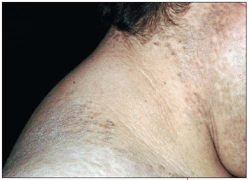
Case 1:
This obese 18-year-old has had a brown, scaly rash for 4 years. The rash spread from his neck, where it initially developed, to his chest and back. It is asymptomatic but of significant concern. His obesity has been an issue since early childhood. A skin biopsy specimen showed papillomatosis, acanthosis, and increased melanin. The rash’s morphological similarity to acanthosis nigricans and the history of obesity suggest confluent and reticulated papillomatosis.
From researchers’ attempts to define the cause of confluent and reticulated papillomatosis, the following basic themes have emerged:
• Endocrine-the association with obesity and puberty and the morphological similarity of the rash to acanthosis nigricans suggests diabetes as the cause.
• Musculoskeletal-the nature of the primary papules suggests a disturbance of keratinization.
• Infectious-the location and morphology of the rash suggest an exaggerated response to Pityrosporum ovale, the infectious organism responsible for tinea versicolor.
NEXT CASE »
For the discussion, click here.
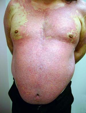
Case 2:
This obese patient presented with widespread, almost confluent psoriasis with intense itching. His obesity suggests concomitant metabolic syndrome. Appropriate blood tests include fasting glucose, hemoglobin A1c, triglycerides, and cholesterol. About 80% of psoriasis cases can be managed with topical medication, but the widespread nature in this case precludes topical therapy. A biologic drug would be a good choice.
Case courtesy of Ted Rosen, MD
NEXT CASE »
For the discussion, click here.
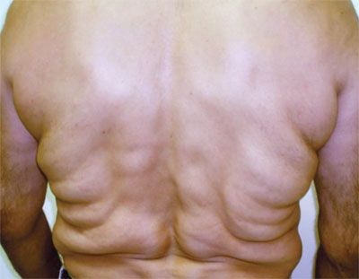
Case 3:
The multiple, symmetrically distributed, soft, nontender swellings on the shoulders and torso of a 56-year-old Hispanic man are characteristic of multiple symmetric lipomatosis (MSL). The painless unencapsulated lipomas are most often located in the subcutaneous tissue of the cervical, deltoid, thoracic, abdominal, and lumbar regions. Because of the symmetry of the masses, the condition is often mistaken for obesity.
Excessive ethanol intake, especially red wine, is often associated with MSL. Other associations are increased blood pressure, dyslipidemia, gout, and decreased insulin sensitivity leading to impaired glucose tolerance or sometimes frank diabetes. MSL must be distinguished from other forms of fatty tumor deposition in the subcutaneous tissues. Of these, Dercum disease, which primarily affects obese postmenopausal women and consists of painful fatty tumors, is the most important.
Case courtesy of Umer Feroze Malik, MD and Sheela Kapre, MD
NEXT CASE »
For the discussion, click here.
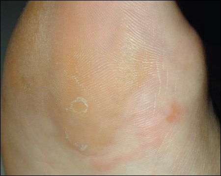
Case 4:
The patient has hyperkeratosis, one of the most common disorders of the feet and particularly common in obese persons. Mechanical forces and hereditary factors contribute to its development. Thickening of the outermost layer of the epidermis occurs over sites that experience increased pressure or friction. Urea 40% cream is helpful.
Image courtesy of Noah S. Scheinfeld, MD, JD
NEXT CASE »
For the discussion, click here.
Case 5:
A 34-year-old morbidly obese man had multiple lesions on both feet that appeared rather rapidly. A review of systems disclosed polyphagia, polydipsia, and polyuria. About 70% to 80% of patients with his condition have diabetes mellitus, although it develops in only 0.3% of patients with diabetes. The lesions were composed of yellowish, firm plaques with large telangiectasia coursing over the top. There was no associated scaling. This is the typical morphology for necrobiosis lipoidica, which favors the feet and forelegs. Potent topical or intralesional corticosteroids may make the lesions disappear. Refractory plaques may respond to oral administration of pentoxifylline.
Case courtesy of Ted Rosen, MD
NEXT CASE »
For the discussion, click here.
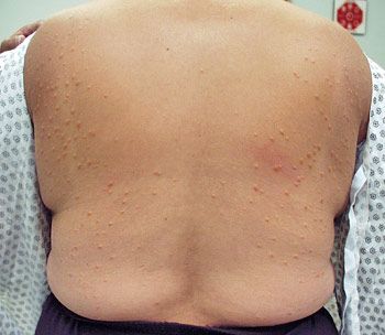
Case 6:
A 57-year-old obese woman with known and poorly controlled type 2 diabetes mellitus presented with the sudden onset of “yellow bumps all over.” Representative lesions on the back are shown. This history and clinical picture are nearly pathognomonic for eruptive xanthomas. Such lesions typically erupt as crops of small, red-yellow papules, most often on the buttocks, shoulders, arms, and legs and may be tender or pruritic. Xanthomas are a sign of primary or secondary hypertrigliceridemia.
If the patient is not already known to have diabetes, glucose intolerance should be strongly suspected and investigated, because most cases of xanthoma are seen in conjunction with diabetes.
Case courtesy of Ted Rosen, MD
