- Clinical Technology
- Adult Immunization
- Hepatology
- Pediatric Immunization
- Screening
- Psychiatry
- Allergy
- Women's Health
- Cardiology
- Pediatrics
- Dermatology
- Endocrinology
- Pain Management
- Gastroenterology
- Infectious Disease
- Obesity Medicine
- Rheumatology
- Nephrology
- Neurology
- Pulmonology
Incidental Finding in Man Referred for Dental Treatment of Snoring
A 62-year-old man with a persistent snoring problem presented for an oral medicine consultation. There was no history of jaw trauma or jaw fracture; jaw function was normal and the patient was not in pain.
A 62-year-old man with a persistent snoring problem presented for an oral medicine consultation. The snoring was disrupting his sleep and that of his partner. He had previously consulted with his dentist and his primary care physician. He had no history of complaints related to jaw function, and he was not in pain. There was no history of jaw trauma or jaw fracture.
Medical History
The patient was healthy, fit, and of normal height and weight. He was taking no medication. A review of systems was unremarkable.
Clinical Findings
Examination revealed very slight facial asymmetry. Mandibular range of motion (ROM) was 41.5 mm. Lateral movement was normal to the right at 9 mm, and slightly reduced to the left at 7 mm. There was audible crepitus in the left temporomandibular joint (TMJ) when the patient opened his jaw. There was no pain with ROM or joint loading. The patient perceived his bite to be normal, and there was positive occlusal contact on the right and left posterior teeth.
Laboratory Findings
There were no laboratory or biopsy findings to report.
Radiology
In patients with snoring who do not have a diagnosis of sleep apnea and are candidates for intra-oral appliance therapy, a panoramic radiograph is required to rule out the possibility of TMJ disease. TMJ disease may be aggravated by use of the appliance or could compromise therapy. The crepitus identified in the patient’s left TMJ required the panoramic study to rule out significant TMJ osteoarthrosis.
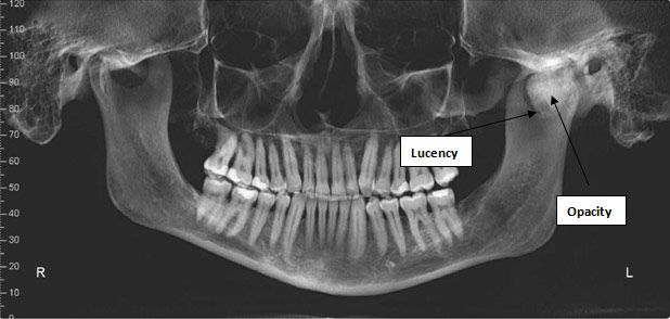
The panoramic radiograph revealed a large irregularly shaped opacity associated with the left mandibular condyle (Figure 1). At the inferior margin there appeared to be a small oval radiolucency. The anterior margin of the opacity was sharply defined, but the superior margin appeared to include an irregular surface contour. The mandible itself was slightly asymmetric, with the height of the left ramus appearing longer than the left. The dentition and bone otherwise appeared normal.

Figure 1
.
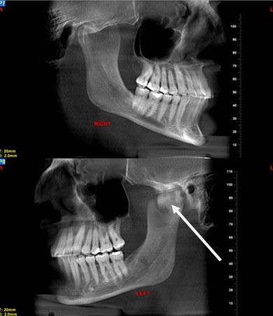
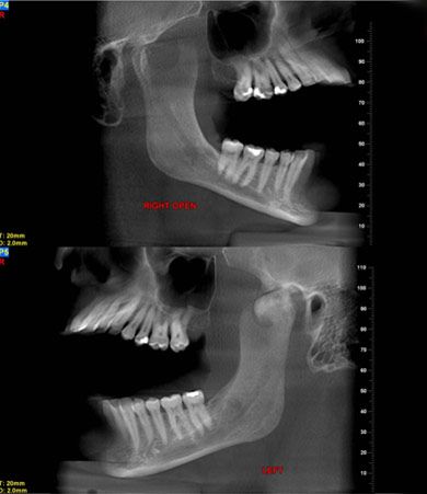
A lateral head/jaw series in the closed and open views (Figure 2) showed a normal jaw opening associated with the abnormality.


Figure 2.
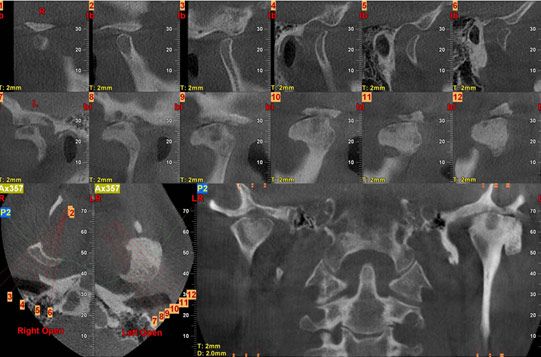
A sagittal series of tomograms (Figure 3) demonstrated a variable opacity with well-formed margins in the left condyle. There was a small ovoid lucency extending through the structure. The head of the mass appeared to extend anteriorly well beyond the fossa.

Figure 3
.
Differential Diagnosis
These findings suggested the following diagnostic possibilities:
• Osteochondroma
• Localized osteochondromatosis
• TMJ dysfunction/TMJ arthritis
Final Diagnosis: TMJ osteochondroma.
Based on the relative symmetry of the condylar mass and cortical surface, the final diagnosis was TMJ osteochondroma. Localized osteochondromatosis is an inherited autosomal dominant condition and typically involves multiple skeletal lesions, which were not observed in this patient. The lesion seen on the radiograph (a large exophytic mass communicating with the condyle) is not consistent with a diagnosis of TMJ dysfunction or TMJ arthritis.
Intervention/Response
Given the absence of functional abnormality or symptoms, no intervention was considered necessary. The condylar finding was incidental in this case and, in the context of normal function, did not preclude use of an intra-oral appliance to help manage the patient’s snoring.
Discussion
Osteochondromas involving the coronoid process or TMJ are slow-growing, asymptomatic, and benign. However, they may be associated with functional disturbance.1-4
Only 38 cases of TMJ osteochondroma have been described in the literature. The findings associated with these cases include mandibular deviation with opening, facial asymmetry, a palpable hard mass in the region of the TMJ, opening limitation, and malocclusion. The mean age of patients in these reported cases is 39.7 years, with pathology found primarily in the fourth decade. This is in contrast to similar tumors involving the axial skeleton, which are reported to occur at an earlier age.
One report4 describes joint ankylosis associated with a concurrent osteochondroma of the mandibular condyle and ipsilateral cranial base. Another case report5 involving a 44-year-old woman describes limited mouth opening and left cheek “swelling.”
The diagnosis is based on clinical symptoms-or lack thereof-and the radiologic findings. In cases of tumor excision, histopathologic evaluation will reveal proliferative bony and hyalinized cartilage-like tissues with a cartilage cap. Surgery is typically unnecessary unless there is concern regarding neoplasm (which has not been reported in association with the TMJ) or severe functional abnormality.6
Teaching Points:
• Facial asymmetry and functional jaw disturbance that is non-painful may be associated with benign non-arthritic disease involving the condyle or coronoid process.
• Osteochondroma is a rare condition but should be considered in the differential diagnosis of a patient with asymptomatic functional jaw problems.
Acknowledgement: Dr Burgess wishes to thank Peter van der Ven, DDS, MSD, PhD, for his gracious contribution of this case.
References:
References:
1. Ribas M de O, Martins WD, de Sousa MH, et al. Osteochondroma of the mandibular condyle: literature review and report of a case. J Contemp Dent Pract. 2007;8:52-59.
2. Henry CH, Granite EL, Rafetto LK. Osteochondroma of the mandibular condyle: report of a case and review of the literature. J Oral Maxillofac Surg. 1992;50:1102-1108.
3. Ord RA, Warburton G, Caccamese JF. Osteochondroma of the condyle: review of 8 cases. Int J Oral Maxillofac Surg. 2010;39:523-528.
4. Karras SC, Wolford LM, Cottrell DA. Concurrent osteochondroma of the mandibular condyle and ipsilateral cranial base resulting in temperomandibular joint ankylosis: report of a case and review of the literature. J Oral Maxillofac Surg. 1996;54:640-646.
5. Saito T, Utsunomiya T, Furutani M, Yamamoto H. Osteochondroma of the mandibular condyle: a case report and review of the literature. J Oral Sci. 2010;4:293-297.
6. Koga M, Toyofuku S, Nakamura Y, et al. Osteochondroma in the mandibular condyle that caused facial asymmetry: a case report. Cranio. 2006;24:67-70.
